Lupine Publishers|Environmental Journals
In this study, we demonstrated the early stage wheat rust diagnosis
using Atomic Force Microscopy (AFM). Wheat rust is a
common disease, caused by parasitic fungus and reduces crop yield up to
30 to 40 percent by effecting leaf and stem of plant. The
wheat seed having fungus are the carrier of rust infection for the
plant. The infected seed and rust fungus not only reduce food
quantity but effect its quality. In our experimental research, the wheat
leaf sample of day 10, 27, 35 and 45 were analyzed. The
rust appears after day 24 on leaf, and rusted leaf has higher surface
roughness than the normal one. We analyzed shape, surface
structure of normal and rusted wheat leaf surface with AFM. The leaf
protein and structure with high-resolution AFM imaging under
controlled environmental conditions. The results revealed the
morphological details of normal and viral infected proteins of wheat
leaf with high-resolution in vivo study. The findings provide the basis
for AFM as a useful tool for investigating microbial-surface
structures and properties at an early stage of rust. We can visualize
the starch granule surface at different stages of maturity reveals
information regarding the development of granule architecture. The
changes in infected leaf compared to normal can be seen at
starch granule interior throughout the growth. Leaf surface shows
depressions on the granule surface and changes in protein for the
infected, and we can manage the disease at an early stage of rust
development. These results will be used for an early stage detection
of wheat rust and can be extended to other crops diseases detection
using AFM and laser-scanning microscopy.
Keywords:Atomic Force Microscopy (AFM); Wheat Rust; Fungal Infection; viral detection
In recent decades, there has been impressive growth in
food production worldwide, which have been attributed to the
development of improved, disease-resistant varieties, increased
use of chemical fertilizers and pesticides. Although enough food is
produced, but yet this food and the technology to produce it does
not match the requirements and as a result about thousand million
people do not get enough to eat and several millions die worldwide
from hunger or hunger-related diseases [1-3]. During the period
1995 to 2050, the world’s population is projected to increase by
75 percent and food security projected to become more critical,
increasing wheat yield potential in the developing world remains
a high priority [4]. Among the other fungal and viral infections,
wheat leaf rust caused by viral fungus continue to pose a major
threat to wheat production over large areas, particularly in Asia.
Rust diseases significantly influence several crop species and
considerable research focuses on understanding the basis of host
specificity and resistance. Like many pathogens, rust fungi vary
considerably in the number of hosts they can infect, such as wheat
leaf rust (Puccinia triticina), which can only infect species in the
genera Triticum and Aegilops. Rusts often produce spots similar to
leaf spots and are bright yellow, orange-red, reddish-brown or black
in color. The pustules are usually raised above the leaf surface, and
some types of rust occur on stems. Rusts are common on grains and
grasses [5-7].
Wheat rusts spread rapidly over long distances by wind. If not detected and treated on time. For effective integrated management of wheat rust diseases close monitoring, international collaboration and strengthening of national capacities are crucial. Although in certain cases fungicide application may be necessary, preventive approaches are the most effective and environmentally friendly means of wheat rust management. specialists from international institutions and wheat producing countries work together to stop these diseases that involves continuous surveillance, sharing data and building emergency response plans to protect their farmers and those in neighboring countries. In general, a fungal infection can cause local or extensive necrosis and can inhibit normal growth of entire plant [8-10].
Several studies worldwide have been carried out for early rust detection. We have applied the surface morphological structure characterization using atomic force microscopy (AFM). It is a non-invasive method and are used for a variety of materials in surface science, biochemistry and biology [11-12]. It is a powerful technique and has the ability to obtain topographic information on, and surface morphology of, the sample. It can also use to investigate chromosomes, proteins, living cells, carbohydrates and DNA [13]. In addition to obtain detailed structural information on the sample and allows us to visualize the cell surface properties on the nanometer scale [14]. Atomic force microscopy (AFM) provides images of biological structures without requiring labeling and to follow dynamic processes in real time. In structural biology, it has proven its ability to image proteins and protein conformational changes at sub molecular resolution, and in proteomics, it is developing as a tool to map surface proteomes and to study protein function by force spectroscopy methods. The power of AFM to combine studies of protein form and protein function enables bridging various research fields to come to a comprehensive, molecular level picture of biological processes [15]. In this study, a wheat leaf rust through the field experiment by the identification and disease index inversion is investigated successfully at an early stage with AFM. The aim of this study is to provide a method for monitoring and evaluating the diseases, so that proper management for rust protection can be made will in time.
The normal and rusted wheat leaf were collected during the
season from National Agricultural Research Council (NARC). The
samples were fixed, and changes are observed using the Atomic
Force Microscopy (AFM, Alpha Contac, Germany). About 40 samples
including 10 control and 30 rusted are taken from field. The AFM
system installed at National Institute of lasers and Optronics
(NILOP) used to analyze the fresh sample taken from field in
1 to 2 hours. The sample of day 10, 27, 35 and 45 are observed.
The rust appears after day 24 on leaf, and rusted leaf has higher
surface roughness than the normal one. The surface imaging was
performed in the contact mode in air. Silicon nitride tips (Alpha
Contac, Germany) were used for all AFM experiments. The radius of
the cantilever of the tip was 7 nm and the diameter of the tip was 14
nm. The length of the cantilever was 12.6 μm, thick-ness 3.52-4.08
μm, width 30-31 μm, and had an oscillation frequency of 287-336
kHz and a force constant of 28-45 N m-1. The images were analyzed
by using WSXM 4.0 Develop 12.1 software for gaining information
from the topography of the cells. The observation was performed
inside a chamber at room temperature [1,2,3,16].
The leaf rust, caused by Puccinia triticina, is an important
disease in most wheat growing areas. The use of genetic resistance
is the most economical and environmentally friendly way to
combat this disease. For early stage rust fungus detection, several
conventional methods are being utilized [17-18]. Atomic force
microscopy is a powerful technique, which allows surface imaging
of non- conducting samples in nanometer scale. In this experiment
wheat leaf are imaged under ambient conditions, i.e. in air and
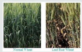
with minimal sample preparations. The wheat corps of normal and
rusted field are shown in Figure 1. Determining the time and scale of
primary infections is also difficult with leaf rust because P. triticinainfected
wheat crops show weak symptoms during the latent
period of the disease. The samples were collected from the field
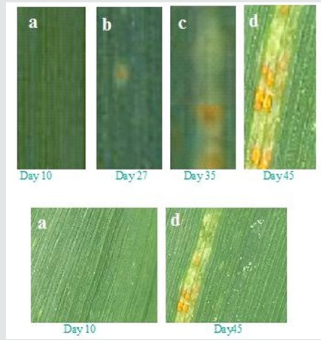
for day 10, 27, 35 and 45 and shown in Figure 2. The rust appears
after day 24 on leaf, and rusted leaf has higher surface roughness
than the normal one Leaf rust symptoms are also checked several
times to distinguish it by stem rust outbreaks at different time.
Wheat leaf rust samples collection and examination of signs and
symptoms in the field is very essential before AFM test. In some
cases, diagnosis of leaf rust often requires isolation of the fungus
and identification of the fungal pathotypes on differential host
genotypes, which is complicated and time-consuming. Monitoring
and early detection of this disease is crucial for the effective control
and implementation of measures. Recent developments of remote
sensing technology had the potential to enable direct detection of
plant diseases under field conditions. However, sometime due to
poor resolution the detection probability reduces.
The cross-section analysis of phase images, the stiffness of granule surfaces for normal and rusted leaf is different. The phase image further visualized the texture of surface depressions, which may be hidden in the topography, and the presence of deep gaps dividing bundles of nodules from each other. In contrast to the surface with blocklets commonly observed and reported in literature [19]. We can see that the presence of 4.5 nm deep depressions with higher stiffness similar to regular granule surface in the background of the amorphous surface suggests that these depressions likely extend to the underneath semi-crystalline growth ring. These depressions might also be a part of internal channels. The wheat tissue morphology can be described as a succession of more or less thick layers including the starchy endosperm layer, the protein stored as granules in the cells of plant seeds layer and the internal cells layers with external pericarp.
In past some researchers worked on an optical light detection for visible and near-infrared region to detect different types of rust at the leaf scale [20]. In this study, none of these indices were able to detect and discriminate the types of rust. However, the anthocyanin reflectance index can be used to detect yellow rust, and the transformed chlorophyll absorption and reflectance index can be used to detect leaf rust [21]. In another study conducted by Frank and Menz, hyperspectral and leaf multispectral data were used to estimate the severity of wheat leaf rust [22]. Results indicated that leaf rust could be detected in the early symptoms by using hyperspectral data. The algorithm used in this research was based on the minimum noise fraction transformation. The reflectance spectra of the infected, non-infected, and dry area, as well as the soil class were taken at the canopy level. Ashourloo et al. showed that the disease symptoms have a high impact on the infected plant reflectance spectra [23]. This means that as the disease severity increases, so does the collected spectrum variations at a specific disease severity. Results showed that as the disease severity increases, the scattering of the numerical values for all of the indices also increases. For the different amounts of scattering and classification accuracy are not the same and depend on the wavelength of light used. Wheat leaf rust at the leaf scale was studied for two purposes, one is to estimate the reflectance spectra of various disease symptoms; and other is to introduce an index for precise determination of disease severity using the spectral reflectance of leaf [24]. In our previous study we have applied optical detection techniques to several viral infection monitoring using human blood in vivo and in vitro [25-32].
In this, experimental studies we demonstrated that Atomic
Force Microscopy AFM provides a powerful platform for detect
wheat leaf rust fungus at nanoscale level. The main advantages of
AFM are the ability to image and manipulate early stage fungus
infection at nanometer resolution and its operation under a wide
variety of physiological conditions for quantifying the physical
properties of cellular structures and leaf surface molecules
topography and structure morphology. The applied technique
provides early stage detection of fungus pathogen, when it it
cannot be observed visually. So, the protection management
with anti-pathogen chemicals can be made to save the crop from
disease. The development and combination of multiple orthogonal,
yet complimentary, biophysical tools will clearly play a major role
in illuminating a deeper understanding of the complex interplay
between physical and biological information.
Collaborations between the Nano, physical and life sciences will lead to the AFM being used more routinely in studies of fundamental and complex biological processes. Such studies will lead to the understanding of the importance of the physical mechanisms governing fungal infection and its characteristics in biology. The atomic force microscopy (AFM) provides the structural, mechanical strength, topography, and surface morphology of the sample in easy way. The information related to the rust fungus infection at an early stage can facilitate the wheat management planning. AFM qualitative and quantitative imaging of granules and stomata support blocklet structural information of diverse starch systems. This imaging methodology will enhance other early stage rust detection techniques to be utilized for wheat food improvements.
Abstract
Keywords:Atomic Force Microscopy (AFM); Wheat Rust; Fungal Infection; viral detection
Introduction
Wheat rusts spread rapidly over long distances by wind. If not detected and treated on time. For effective integrated management of wheat rust diseases close monitoring, international collaboration and strengthening of national capacities are crucial. Although in certain cases fungicide application may be necessary, preventive approaches are the most effective and environmentally friendly means of wheat rust management. specialists from international institutions and wheat producing countries work together to stop these diseases that involves continuous surveillance, sharing data and building emergency response plans to protect their farmers and those in neighboring countries. In general, a fungal infection can cause local or extensive necrosis and can inhibit normal growth of entire plant [8-10].
Several studies worldwide have been carried out for early rust detection. We have applied the surface morphological structure characterization using atomic force microscopy (AFM). It is a non-invasive method and are used for a variety of materials in surface science, biochemistry and biology [11-12]. It is a powerful technique and has the ability to obtain topographic information on, and surface morphology of, the sample. It can also use to investigate chromosomes, proteins, living cells, carbohydrates and DNA [13]. In addition to obtain detailed structural information on the sample and allows us to visualize the cell surface properties on the nanometer scale [14]. Atomic force microscopy (AFM) provides images of biological structures without requiring labeling and to follow dynamic processes in real time. In structural biology, it has proven its ability to image proteins and protein conformational changes at sub molecular resolution, and in proteomics, it is developing as a tool to map surface proteomes and to study protein function by force spectroscopy methods. The power of AFM to combine studies of protein form and protein function enables bridging various research fields to come to a comprehensive, molecular level picture of biological processes [15]. In this study, a wheat leaf rust through the field experiment by the identification and disease index inversion is investigated successfully at an early stage with AFM. The aim of this study is to provide a method for monitoring and evaluating the diseases, so that proper management for rust protection can be made will in time.
Materials and Methods
Results and Discussion
Figure 2: The wheat rusts fungal infected leaf samples of day 10 (a) normal and for day 27 (b), day 35(c) and day 45(d) are
collected and analyzed with Atomic Force microscopy (AFM) topographic, phase images, and their cross-section analysis.

The AFM study of rusted wheat leaf from very early stage of
growth provides a means to quantify their mechanical properties
and examine their response to nanoscale forces, pulling single
surface proteins with a functionalized tip allow one to understand
their role in sensing and adhesion. The combination of these
nanoscale techniques with modern molecular biology approaches,
genetic engineering and optical microscopies provides a powerful
platform for understanding the sophisticated functions of the plant
machinery, and its role in the onset and progression of complex
diseases. Topographic image and the cross-section analysis of the
samples of day 10, 27, 35 and 45 were imagine with AFM. The
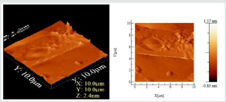
results can be seen in Figure 3 for the sample collected on day
10 (normal sample). It can be seen in Figures 2(a) & 3, no fungus
attack is observed on leaf and it represents normal study. The rust
fungus appears after day 24 on leaf, and rusted leaf has higher
surface roughness than the normal one at day 10. The leaf starch
granule surface structure can be seen in Figure 3. The observation
represents essentially a top view of the surface enabling estimations
in two and three dimensions of the size of different microstructures.
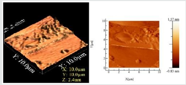
The rust fungus appears after day 24 and the sample in Figure 2(bd)
and two and three-dimensional AFM images in Figure 4. As seen
in the topographic image block lets are clearly visible in image.
Figure 3: The Atomic Force microscopy (AFM) 2D and 3D topographic, phase image of Wheat leaf rust infection at day 10 for
normal leaf. Scale of the axis of the cross-section analysis of topographic is 10 μm, angle 10-15o, whereas depth is 2.4 nm.


Figure 4: The Atomic Force microscopy (AFM) 2D and 3D topographic, phase image of Wheat leaf rust infection at day 45 for
rust fungus infection. Scale of the axis of the cross-section analysis of topograph is 10 μm, angle 10-15o, whereas depth is 2.4
nm.

The AFM 2D and 3D images had dimensions between 10 x10
μm. Apparently, the blocklets were clustered or fused together
at different heights forming nodules and are mostly elongated
in shape. Depressions were also observed on the surface of the
granule. Phase image in an AFM study highlights the stiffness of the
sample surface. It records the phase lagging between the cantilever
oscillation and the phase of driving signal giving an indication of
relative stiffness at different locations of the surface of the sample,
indicating rust formation. Thus, phase images can be utilized
to recognize fungus and understand the microstructure inside
depressions, which were hidden in the topographic image. The
topographic image shows some corresponding features, surface
roughness hinders the identification of domains. The phase image
allows unambiguous resolution of the different material phases.
The surface of the day 35 and 45 are similar to day 27 and indicate
that top of the nodules was mostly stiffer than valleys, as shown in
Figure 4.
The cross-section analysis of phase images, the stiffness of granule surfaces for normal and rusted leaf is different. The phase image further visualized the texture of surface depressions, which may be hidden in the topography, and the presence of deep gaps dividing bundles of nodules from each other. In contrast to the surface with blocklets commonly observed and reported in literature [19]. We can see that the presence of 4.5 nm deep depressions with higher stiffness similar to regular granule surface in the background of the amorphous surface suggests that these depressions likely extend to the underneath semi-crystalline growth ring. These depressions might also be a part of internal channels. The wheat tissue morphology can be described as a succession of more or less thick layers including the starchy endosperm layer, the protein stored as granules in the cells of plant seeds layer and the internal cells layers with external pericarp.
In past some researchers worked on an optical light detection for visible and near-infrared region to detect different types of rust at the leaf scale [20]. In this study, none of these indices were able to detect and discriminate the types of rust. However, the anthocyanin reflectance index can be used to detect yellow rust, and the transformed chlorophyll absorption and reflectance index can be used to detect leaf rust [21]. In another study conducted by Frank and Menz, hyperspectral and leaf multispectral data were used to estimate the severity of wheat leaf rust [22]. Results indicated that leaf rust could be detected in the early symptoms by using hyperspectral data. The algorithm used in this research was based on the minimum noise fraction transformation. The reflectance spectra of the infected, non-infected, and dry area, as well as the soil class were taken at the canopy level. Ashourloo et al. showed that the disease symptoms have a high impact on the infected plant reflectance spectra [23]. This means that as the disease severity increases, so does the collected spectrum variations at a specific disease severity. Results showed that as the disease severity increases, the scattering of the numerical values for all of the indices also increases. For the different amounts of scattering and classification accuracy are not the same and depend on the wavelength of light used. Wheat leaf rust at the leaf scale was studied for two purposes, one is to estimate the reflectance spectra of various disease symptoms; and other is to introduce an index for precise determination of disease severity using the spectral reflectance of leaf [24]. In our previous study we have applied optical detection techniques to several viral infection monitoring using human blood in vivo and in vitro [25-32].
Conclusion
Collaborations between the Nano, physical and life sciences will lead to the AFM being used more routinely in studies of fundamental and complex biological processes. Such studies will lead to the understanding of the importance of the physical mechanisms governing fungal infection and its characteristics in biology. The atomic force microscopy (AFM) provides the structural, mechanical strength, topography, and surface morphology of the sample in easy way. The information related to the rust fungus infection at an early stage can facilitate the wheat management planning. AFM qualitative and quantitative imaging of granules and stomata support blocklet structural information of diverse starch systems. This imaging methodology will enhance other early stage rust detection techniques to be utilized for wheat food improvements.
For more Lupine
Publishers Open Access Journals Please visit our website:
http://lupinepublishers.us/
For more Open Access Journal on Environmental and Soil Sciences articles Please Click Here:
https://lupinepublishers.com/environmental-soil-science-journal/
http://lupinepublishers.us/
For more Open Access Journal on Environmental and Soil Sciences articles Please Click Here:
https://lupinepublishers.com/environmental-soil-science-journal/
To Know More About Open Access Publishers Please Click on Lupine Publishers


No comments:
Post a Comment